Diabetic Foot Treatment
Opening artery blockage with balloon and stent without surgery in diabetic foot ulcers; early assessment to save the foot.
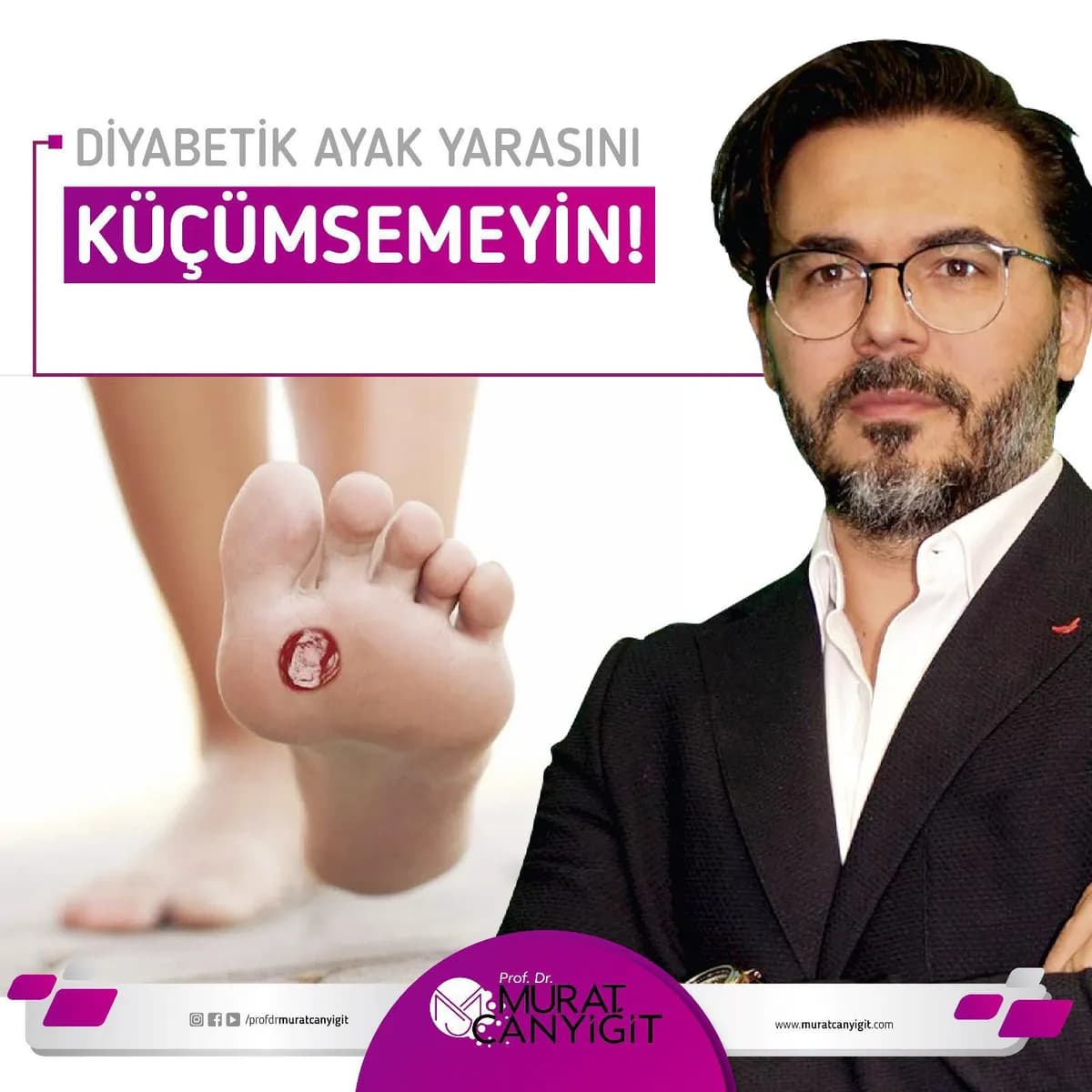
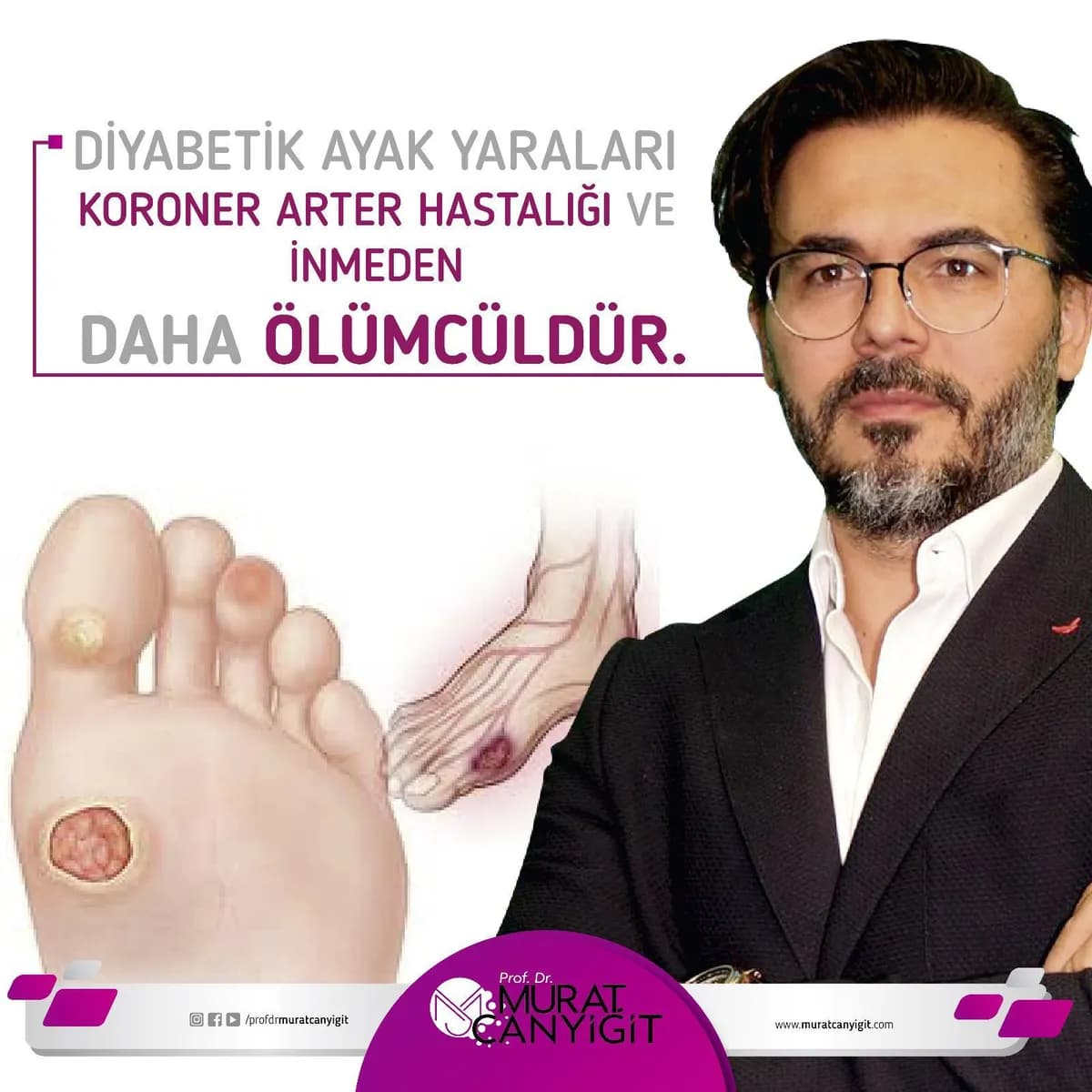
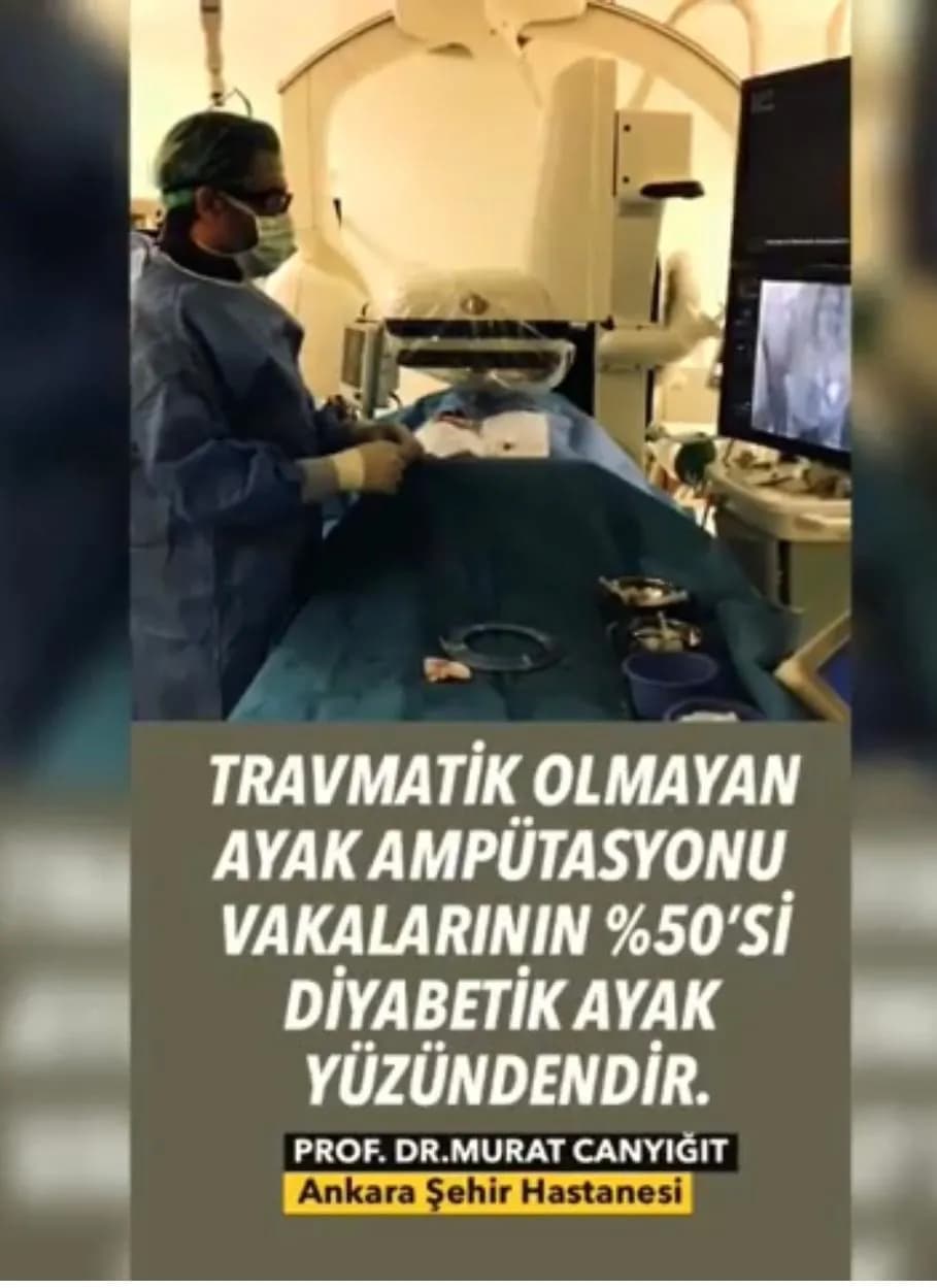
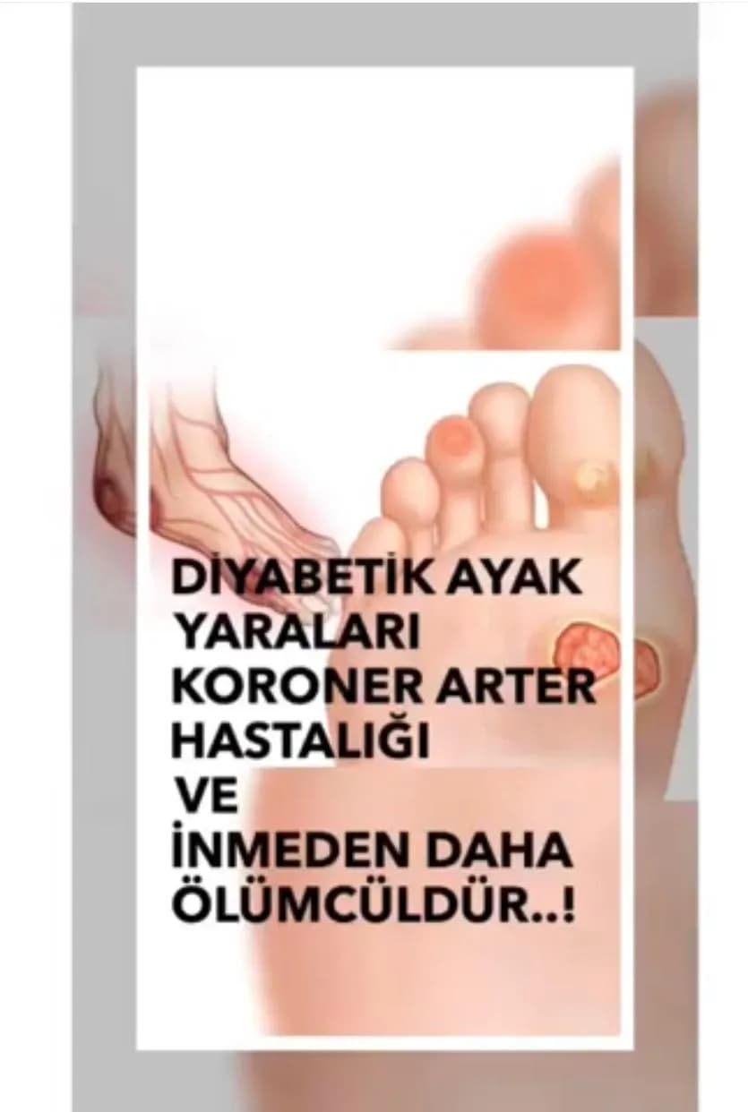
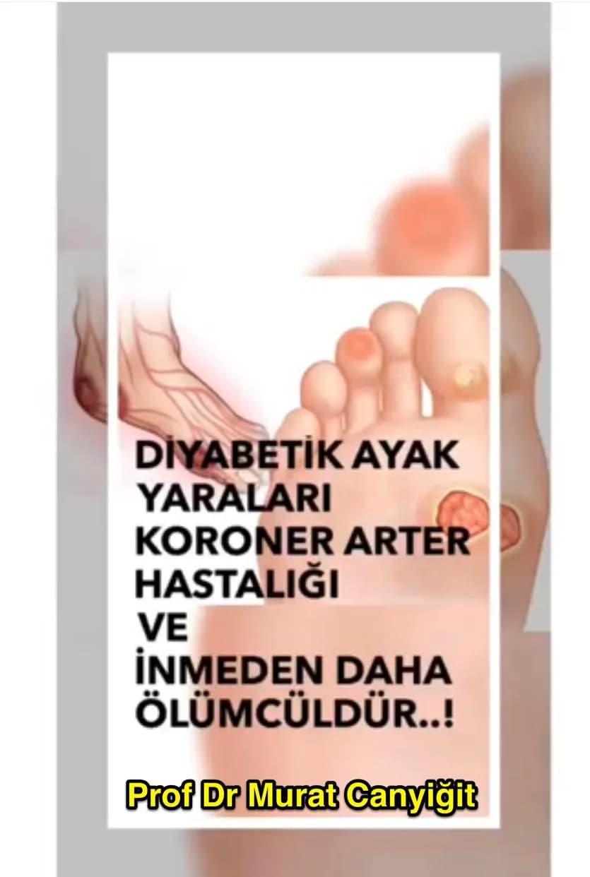
Diabetic foot is the common name for wounds and infections that develop on the feet of people with diabetes. Not everyone with diabetes develops a foot ulcer, but a large share of the ulcers that do develop involve an artery blockage, which is why they do not heal. Diabetic foot is one of the particular areas of interest of Prof. Dr. Murat Canyiğit.
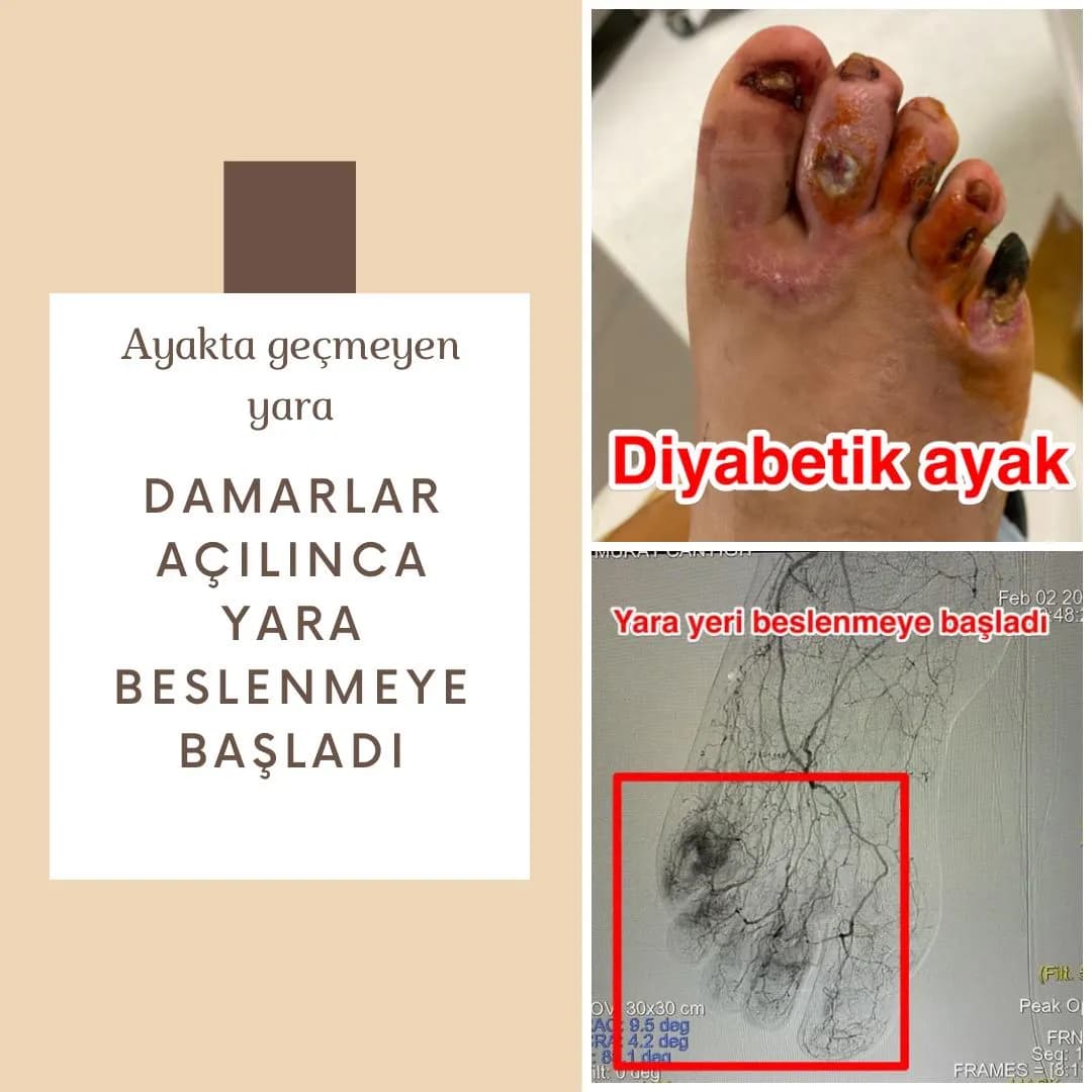
When not enough blood reaches the wound even the best wound care falls short. In interventional radiology a thin catheter introduced at the groin or ankle can open the blocked arteries with a balloon and, if needed, a stent, without surgery. The aim is to support healing of the wound and to save the foot.
Why does a diabetic foot ulcer not heal?
Three factors act together: nerve damage, artery blockage and infection. Nerve damage (neuropathy) causes loss of sensation in the foot, so a small wound, callus or shoe rubbing goes unnoticed. Artery blockage reduces blood flow to the wound, making it harder for the tissue to receive oxygen and immune cells. High blood sugar both predisposes to infection and slows healing.
Treatment is planned according to which of the three dominates. If there is an artery blockage, healing is very difficult until blood flow is restored and the risk of limb loss rises.
Which signs call for prompt assessment?
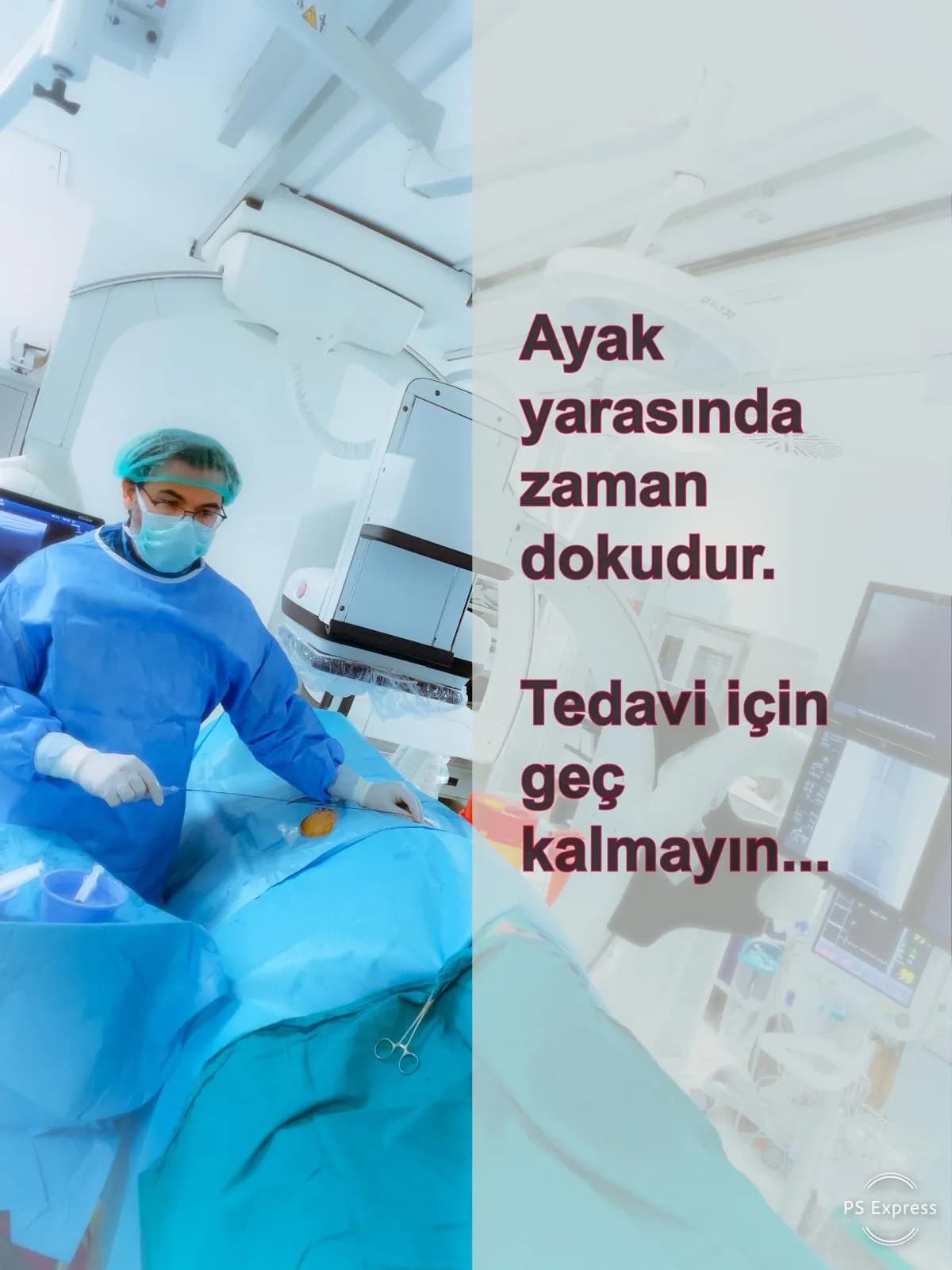
A wound that lasts longer than two to three weeks or keeps growing, bluish or black toes, a cold and pale foot, foot pain at rest and especially at night, calf pain when walking, and redness, swelling or foul-smelling discharge around a wound are important warning signs.
Fever, rapidly spreading redness, blackening or pus from the wound is an emergency; seek care at a healthcare facility without delay.
How are the arteries assessed?
At examination, the foot pulses and the skin are assessed. The ankle-brachial index (ABI) and color Doppler ultrasound are the first steps. In diabetic patients the arteries are calcified so ABI can be falsely high; toe pressure and Doppler waveforms are therefore also used. CT or MR angiography is added if needed. Definitive diagnosis and treatment are often carried out in the same session with catheter angiography.
Interventional radiology treatment: balloon and stent
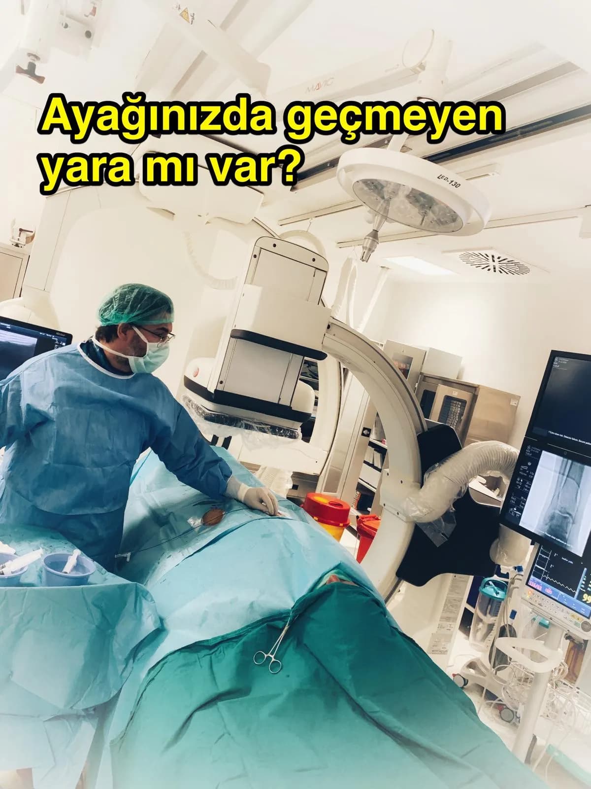
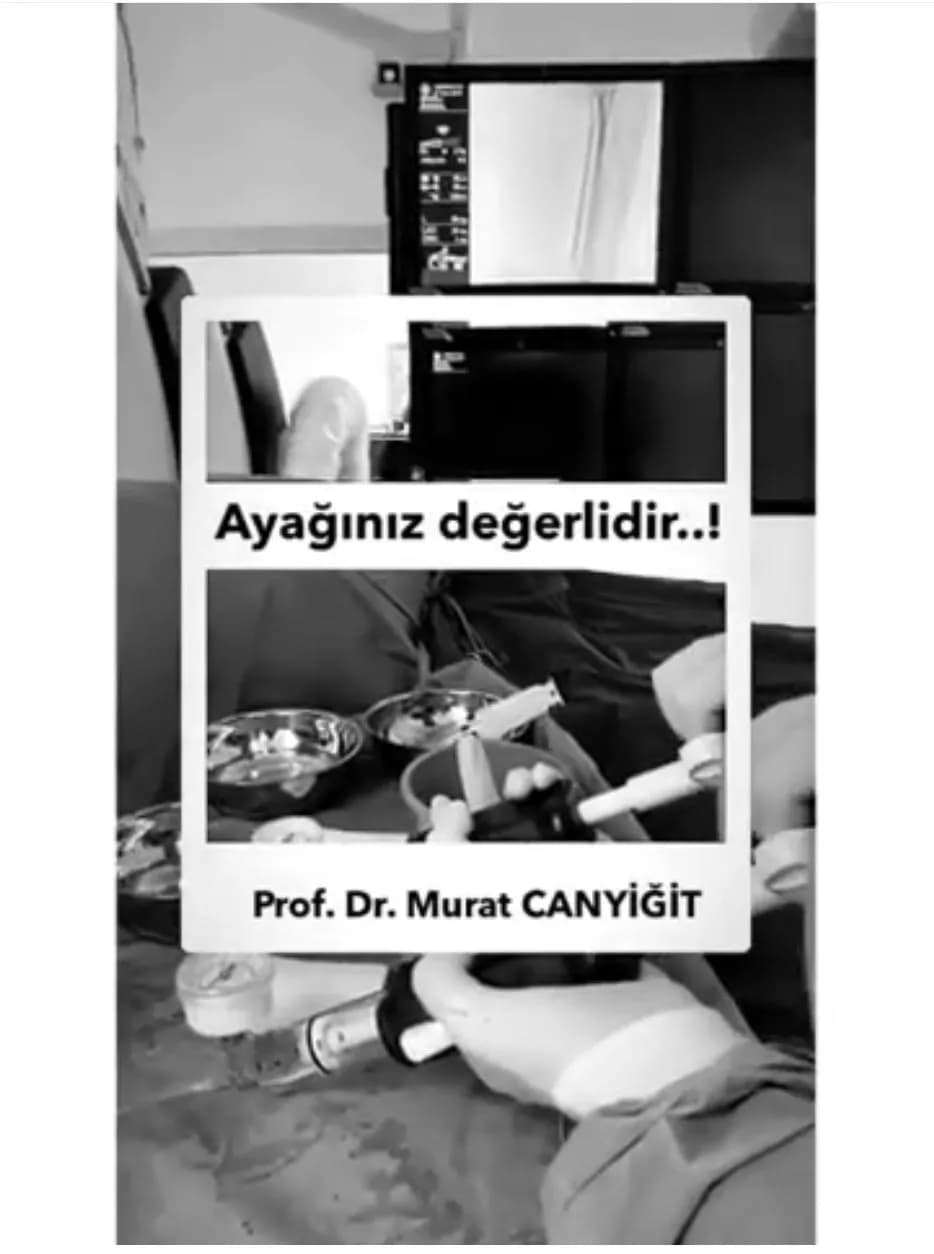
A needle is used to enter the artery at the groin or ankle, a thin catheter is advanced to the blocked segment and the artery is opened with a balloon (angioplasty). If needed a stent is placed to keep it open. The procedure is often done under local anesthesia and requires no large incision. This approach is particularly valuable for patients with a high surgical risk.
In diabetic foot the blockage is most often in the thin arteries below the knee. These can be opened with small-diameter balloons. When the blockage cannot be reached from the groin, it can be crossed from the opposite direction through a vessel at the ankle (transpedal approach). In calcified arteries intravascular ultrasound (IVUS) helps choose the balloon size.
Published experience
In a 2026 study by Prof. Dr. Murat Canyiğit and colleagues, foot ulcers were present in 90% of 40 patients who underwent balloon angioplasty of below-the-knee arteries. Over a mean follow-up of 92 days, wound healing was 69.7% and limb salvage 94.6%; no patient needed target-vessel reintervention. In a 2025 study of IVUS-guided angioplasty in severely calcified below-the-knee arteries a median of only 2 mL contrast was used and no major amputation occurred; wounds healed in 62.5% of those with ulcers.
These are single-center retrospective studies with limited patient numbers and follow-up. They do not mean the same result will be obtained in every patient.
Treatment is teamwork
Opening the artery is only one part of treatment. Wound care, treatment of infection, removal of dead tissue, blood sugar control, special footwear or insoles that offload the foot and, when needed, orthopedic or plastic surgery support are carried out together. The interventional radiologist provides the link that restores blood flow; working together with the diabetes and wound team determines the outcome.
Daily care to protect the foot
Check your feet every day, especially the soles and between the toes, using a mirror. Do not walk barefoot and wear comfortable, wide-toed, seamless shoes. Cut nails straight, do not cut calluses yourself and do not warm the feet with hot-water bottles or heaters; when sensation is reduced a burn may go unnoticed. Keep blood sugar in the target range, stop smoking and keep blood pressure and cholesterol under control.
After the procedure and risks
After the procedure the access site is observed for a short time. Many patients go home the same or the next day. Regular use of the blood thinners recommended by the doctor, wound care and follow-up continue.
Risks include bleeding or bruising at the access site, vessel injury, allergy or kidney effects related to contrast, re-narrowing of the artery and, rarely, the need for a further intervention. Risks vary with the patient's general condition and are discussed in detail before the procedure.
Consultation and appointments
Consultations and procedures are carried out in person at Ankara City Hospital, Neuro-orthopedics Building, Department of Radiology, Interventional Radiology Unit; online consultation is not offered. Please bring previous reports, films and CDs, your current medication list (especially blood thinners and diabetes drugs) and any blood test results. See the Book an Appointment page for details.
Television broadcast
Diabetes and diabetic foot (Günün Raporu) · BT
Information Graphics

FAQ
Will a diabetic foot ulcer heal once the artery is opened?
Opening the artery supports healing by bringing blood to the wound, but the result also depends on wound depth, infection, blood sugar control and wound care. Published studies report healing in a proportion of patients; it is not guaranteed for everyone.
I was told my foot would be amputated: can the endovascular method be tried?
In some patients restoring blood flow can prevent amputation; this decision is made after vascular imaging and examination. If there is infection or extensive tissue death these must be treated first. Please seek a specialist's assessment.
Is the procedure painful and is anesthesia needed?
Local anesthesia is enough in most patients; brief discomfort may be felt at the access site. Light sedation is added if needed.
Can the artery block again?
The artery can narrow again over time. Quitting smoking, regular medication, blood sugar control and regular follow-up reduce this risk. The procedure can be repeated if needed.
Scientific sources
- Canyiğit M, Beşler MS, Kaya T. Physiological indices for evaluating balloon angioplasty outcomes in below-the-knee artery lesions of patients with CLTI. Diagn Interv Radiol, 2026
- Beşler MS, Sözeri A, Canyiğit M. Balloon angioplasty under intravascular ultrasound guidance in calcified below-the-knee tibial arteries. Diagn Interv Radiol, 2025
- Canyiğit M, Ateş ÖF, Beşler MS. Primary retrograde transpedal approach for revascularization of chronic total occlusions of the superficial femoral artery. Diagn Interv Radiol, 2021
The information on this page is general and does not replace an examination. Diagnosis and treatment decisions are made only after an in-person consultation. In an emergency (sudden chest or back pain, signs of stroke, rapidly progressing blackening or infection of the foot) call 112 without waiting.
Book Appointment