Leg Artery Blockage & Diabetic Foot
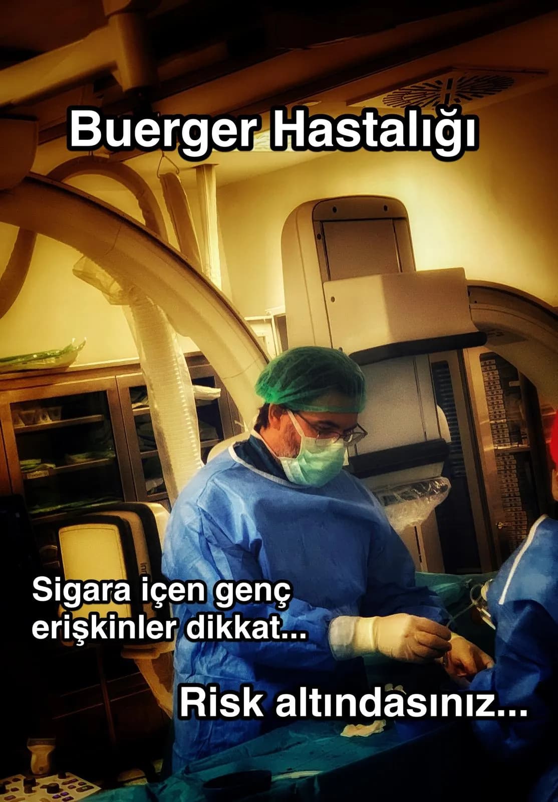
Opening narrowed or blocked leg arteries without open surgery, using balloon and stent.
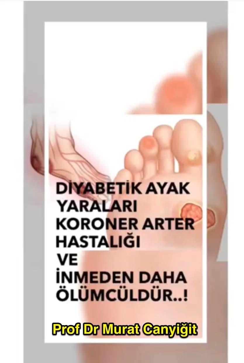
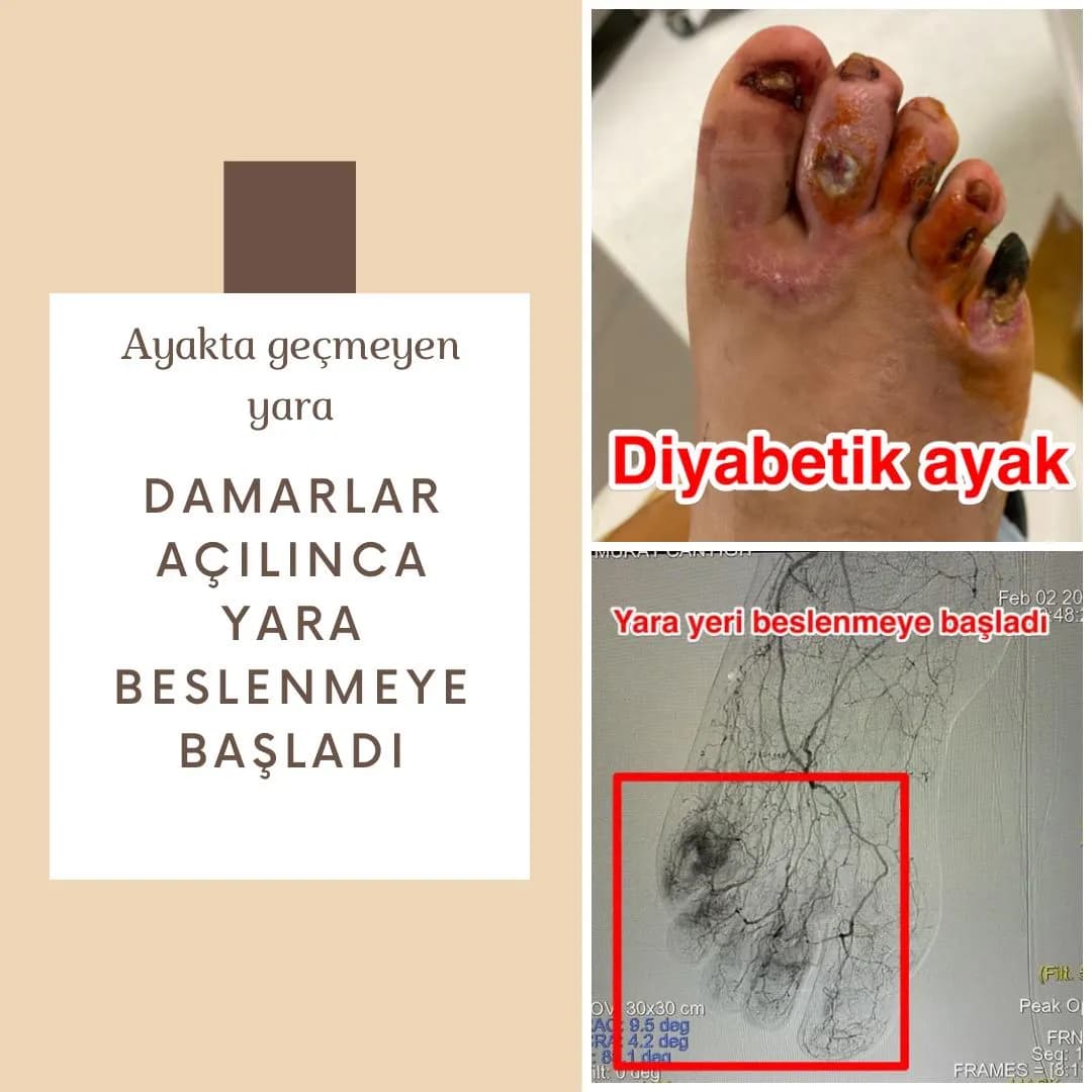
A blockage in the leg arteries may cause pain when walking, cold feet and wounds that do not heal. In diabetic patients, restoring blood flow is critical for wound healing.
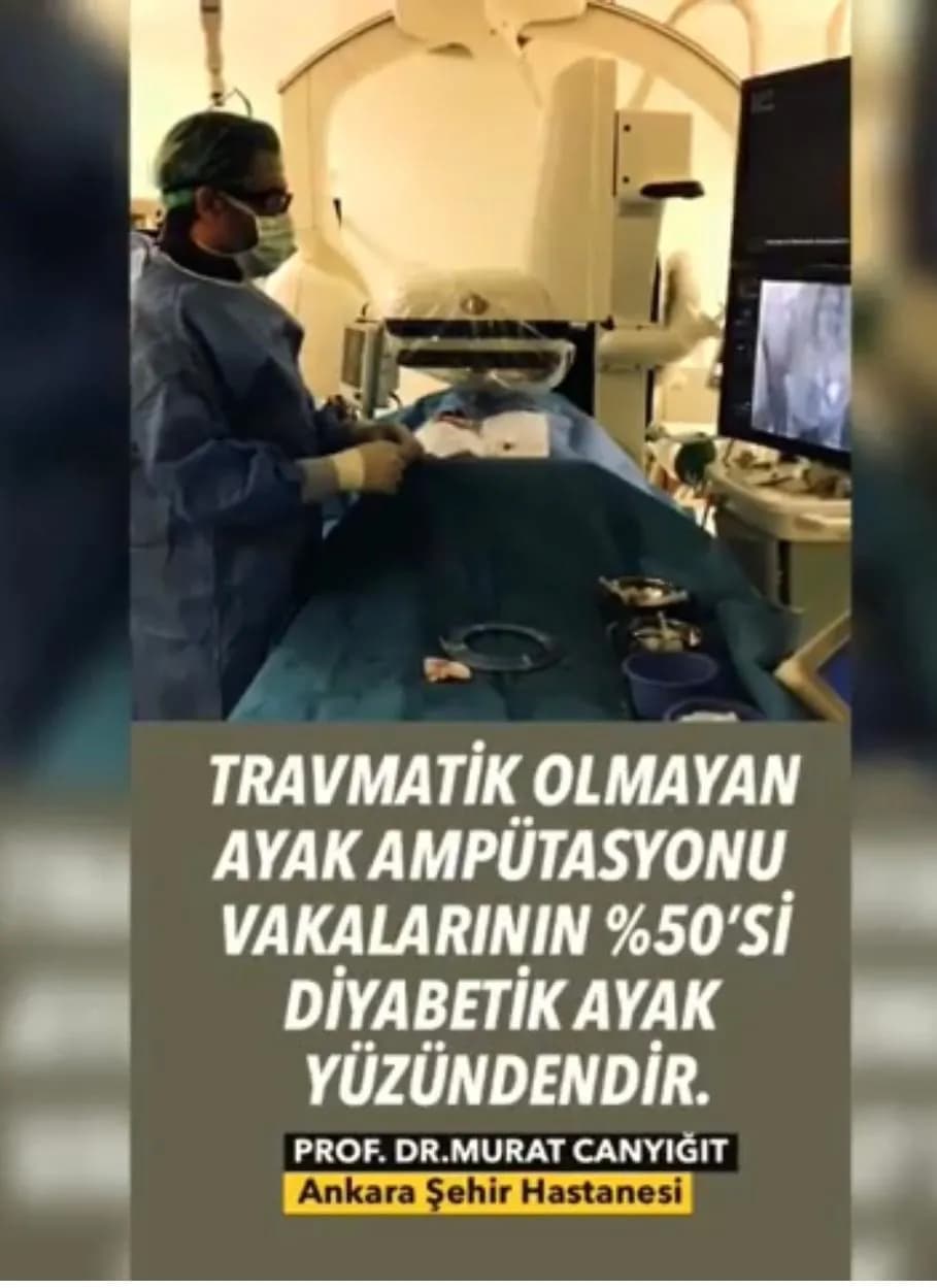
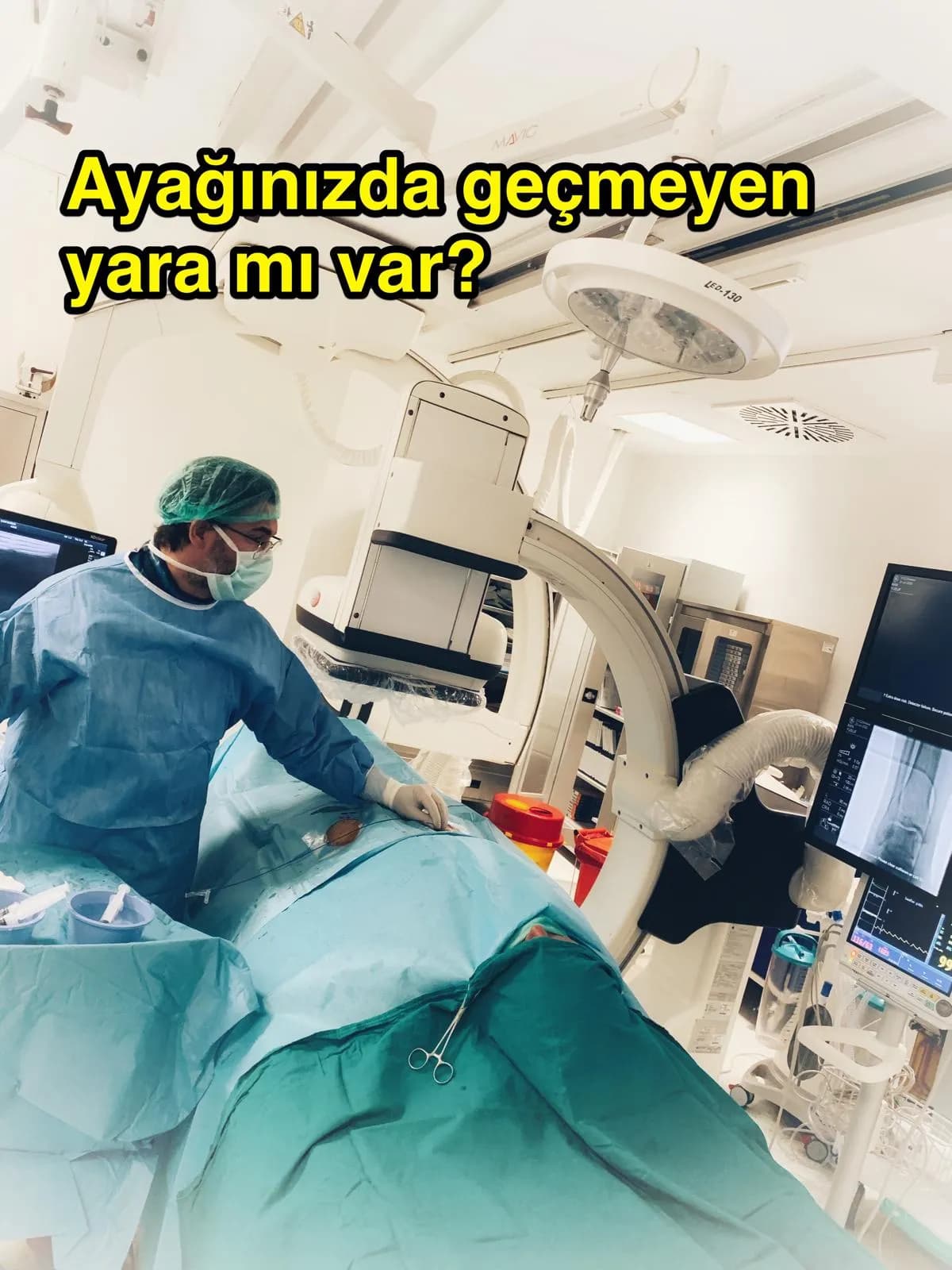
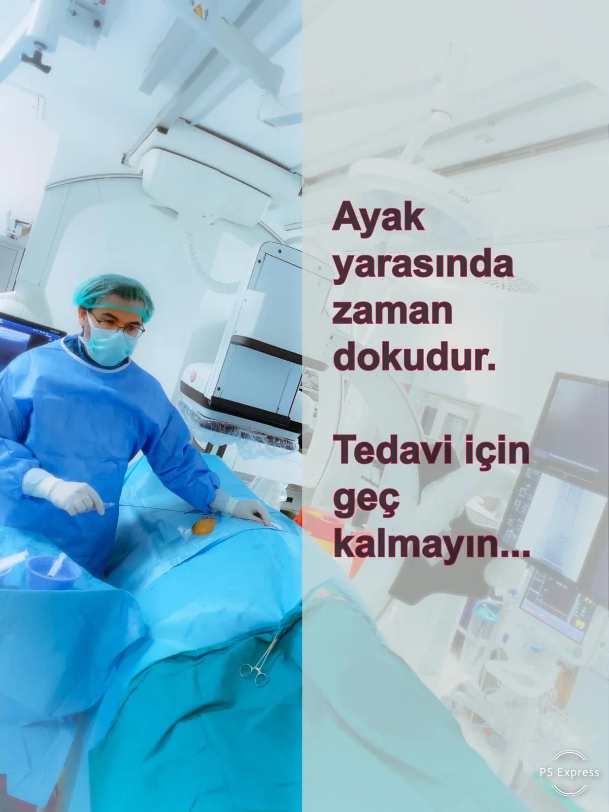
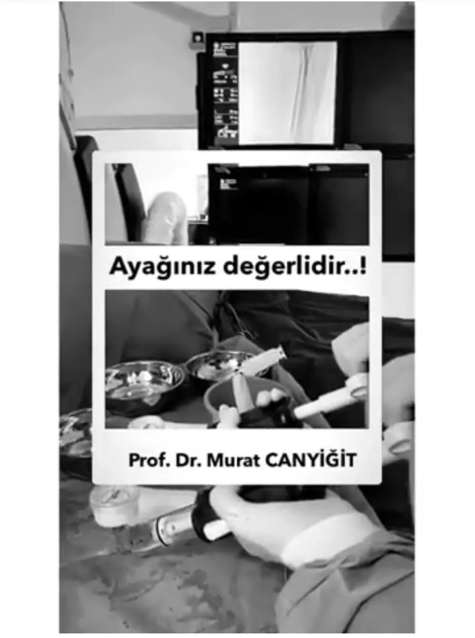
In interventional radiology, a thin catheter is advanced through the groin or ankle to reach the blocked artery, which is then opened with a balloon (angioplasty) and a stent when needed. The procedure is usually done under local anesthesia without a large incision.
Every patient's vessels are different; the right approach is decided after examination and angiography.
What is leg artery blockage?
The arteries that carry blood to the legs can gradually narrow or become completely blocked by cholesterol plaque and calcification. This is called peripheral artery disease. Smoking, diabetes, high blood pressure, high cholesterol and older age are the main risk factors. As the artery narrows, less blood reaches the leg and foot and the tissue does not get enough oxygen.
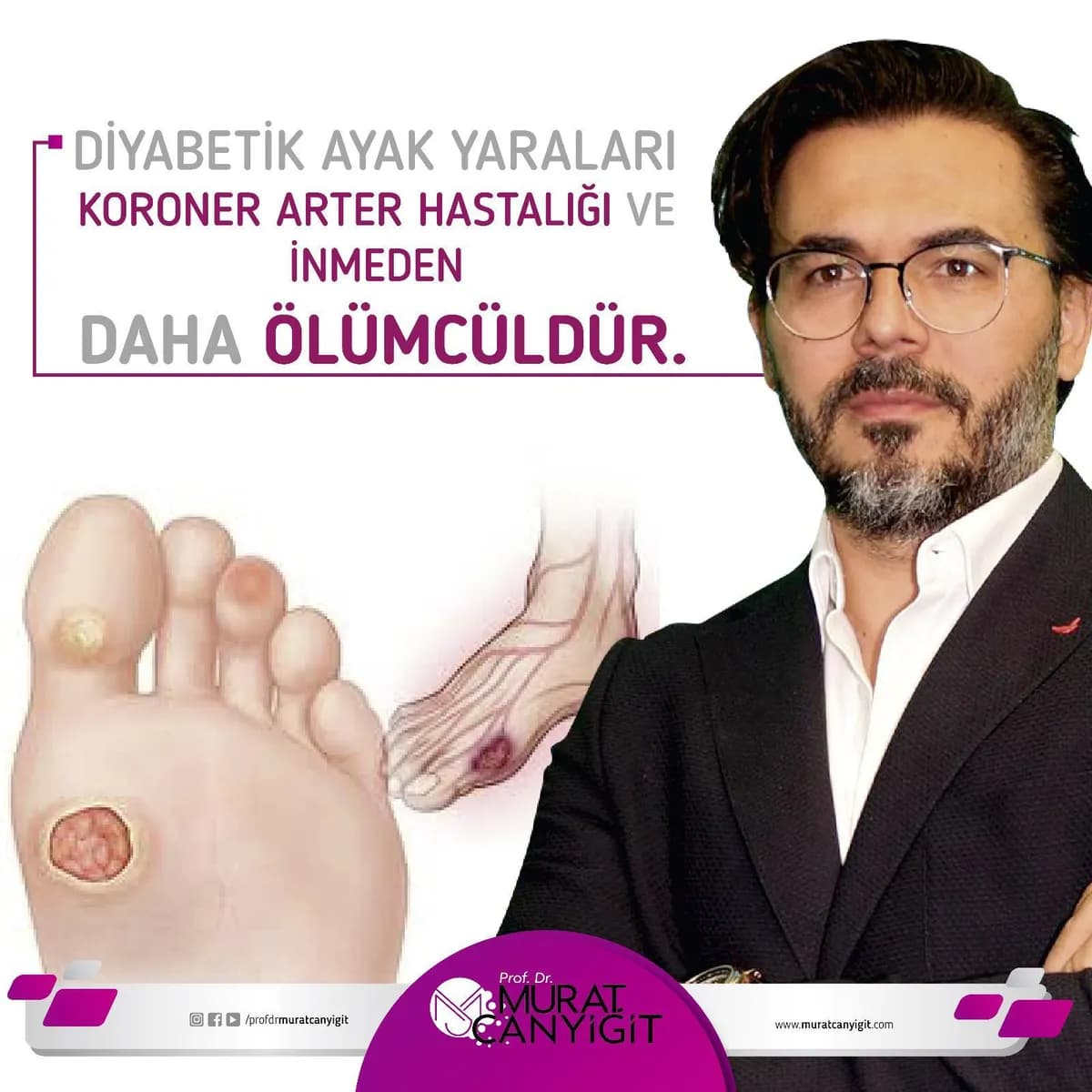
The disease often starts silently. As it progresses it first causes symptoms when walking and later at rest. A wound on the foot that does not heal or darkening of the toes can indicate an advanced stage of blockage and carries a risk of limb loss.
Which symptoms call for assessment?
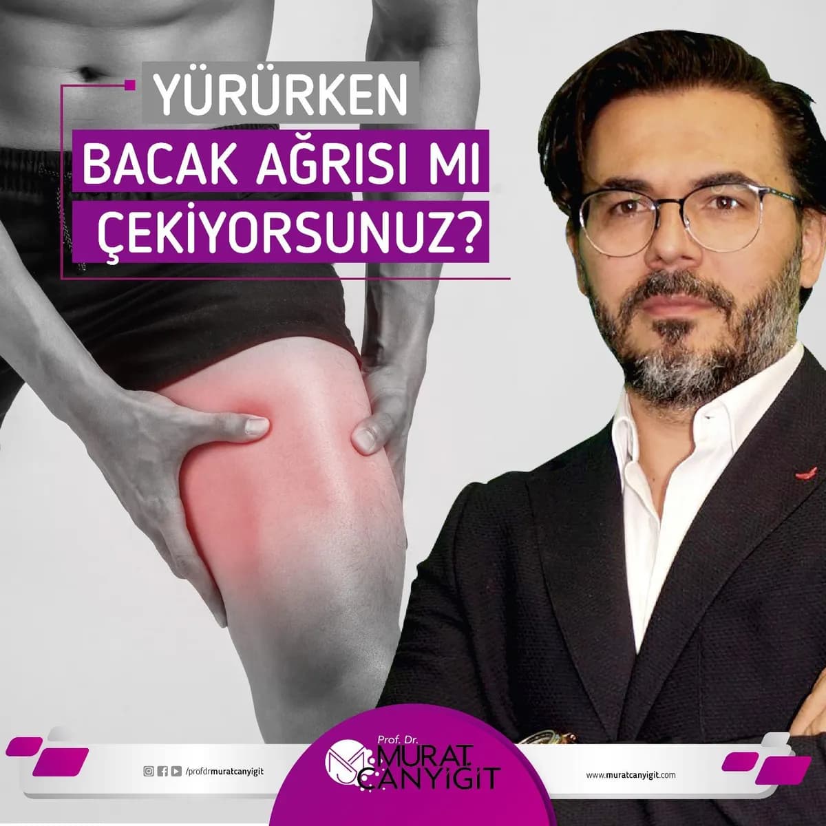
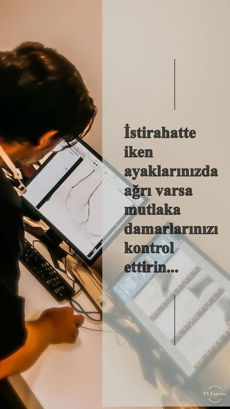
Pain in the calf, thigh or buttock that starts after walking a few hundred meters and eases with rest (claudication) is the most typical first symptom. A cold leg or foot, changes in the nails and skin, hair loss, foot pain at night in bed that eases when the leg hangs down, and a wound or crack that does not heal are other warning signs.
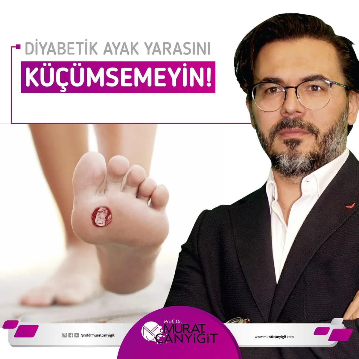
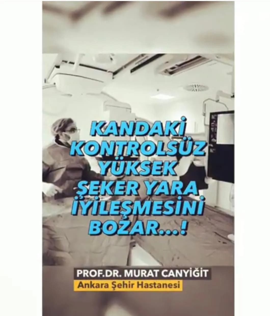
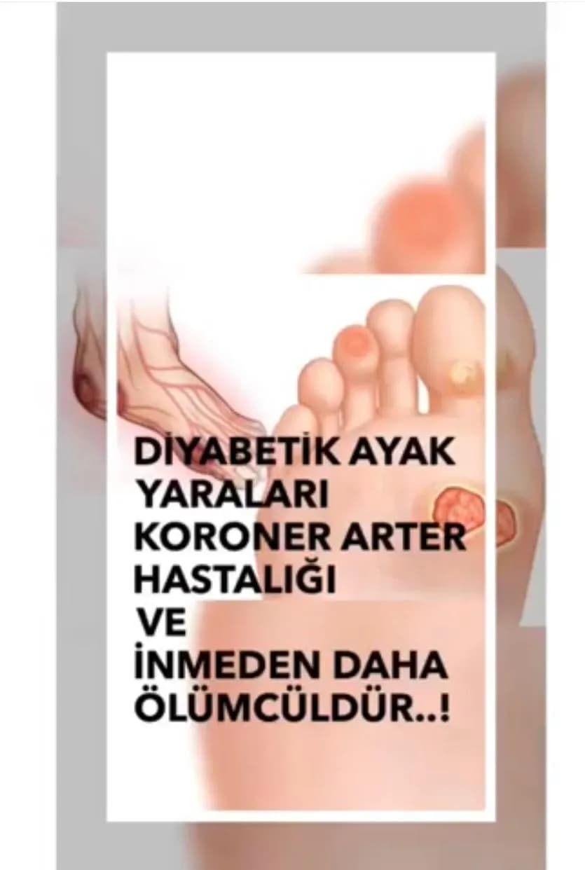
In a diabetic patient with a foot ulcer, the state of the arteries must be assessed in addition to local wound care. Without enough blood flow it is very hard for a wound to heal.
How is it diagnosed?
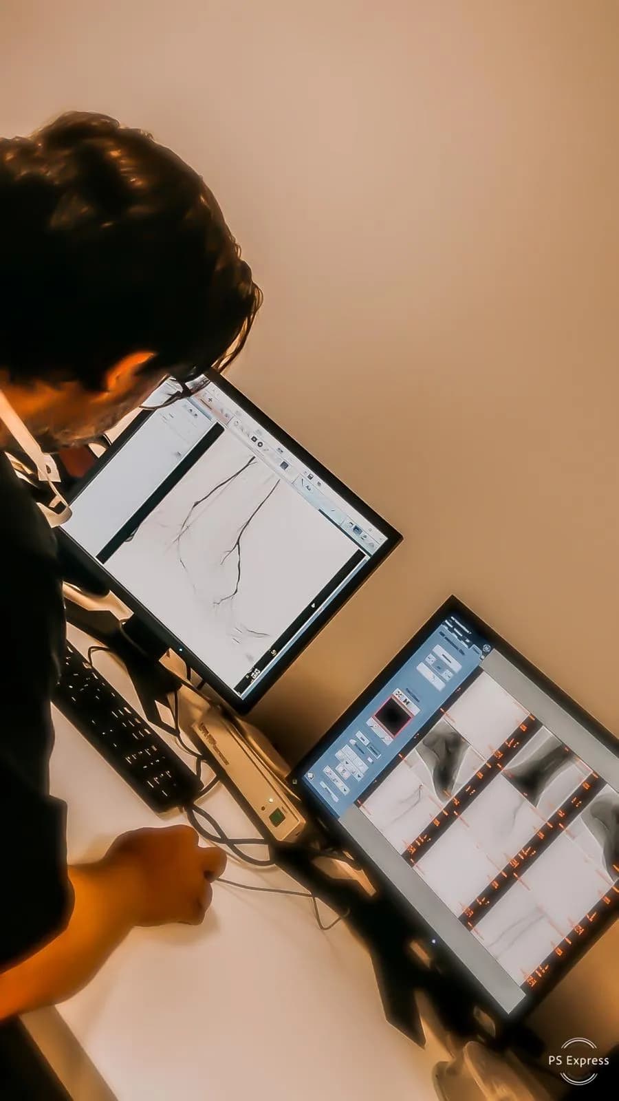
At examination the pulses and the foot are assessed. The ankle-brachial index (ABI) and color Doppler ultrasound are the first steps. If needed, CT or MR angiography is performed. Definitive imaging and treatment can be carried out in the same session with catheter angiography.
Incision-free treatment: balloon and stent
In interventional radiology a needle is used to enter the artery at the groin or ankle, a thin catheter is advanced to the blocked segment and the artery is opened with a balloon (angioplasty). If needed a stent is placed to keep the artery open. It is usually done under local anesthesia and requires no large incision as in surgery.
The thin arteries below the knee can also be opened with small-diameter balloons in suitable patients. When the blockage cannot be reached from the groin, it can be crossed from the opposite direction through a vessel at the ankle (transpedal approach). The route is chosen according to the vessel anatomy.
Published experience
In a 2026 study by Prof. Dr. Murat Canyiğit and colleagues, foot ulcers were present in 90% of 40 patients who underwent balloon angioplasty of below-the-knee arteries. Over a mean follow-up of 92 days wound healing was 69.7% and limb salvage 94.6%. In a 2025 study, calcified below-the-knee arteries were opened under intravascular ultrasound guidance using only a median of 2 mL contrast. In a 2021 study the ankle route was used in 23 patients with chronic total occlusion of the thigh artery; one-year primary patency was 78%.
These are single-center retrospective studies; they do not mean the same result will be obtained in every patient.
After the procedure
After the procedure the access site is observed for a short time. Many patients go home the same or the next day. Taking the blood thinners recommended by the doctor regularly, quitting smoking, keeping blood sugar and cholesterol under control and walking regularly are very important for lasting results. In diabetic foot ulcers wound care and follow-up continue.
Risks
Like any intervention this procedure has risks: bleeding or bruising at the access site, vessel injury, allergy or kidney effects related to contrast, re-narrowing of the artery and, rarely, the need for a further intervention. Risks vary with the patient's general condition and are discussed in detail before the procedure.
What happens at the consultation?
At the first consultation your complaints, medical history, medications (especially blood thinners and diabetes drugs), allergies and previous imaging and test results are reviewed together. Foot pulses, walking distance, any foot wound and diabetes history are assessed in particular. Please bring previous reports, films and CDs, your current medication list and any blood test results.
If needed, additional imaging or tests are requested. Whether the treatment is suitable, how the procedure will be done, the possible risks and the alternatives are explained in detail; your questions are answered and the procedure is planned after your consent is obtained.
Preparation before the procedure
Preparation instructions are given according to the type of procedure and the patient's condition. In general this involves fasting for a set time before the procedure, having tests that show kidney function and bleeding-clotting values and adjusting some medications as advised by the doctor. Always tell us if you are allergic to contrast material or medicines.
Having a relative with you on the day of the procedure is helpful for returning home after discharge.
Where and how to book
Consultations and procedures are carried out in person at Ankara City Hospital, Neuro-orthopedics Building, Department of Radiology, Interventional Radiology Unit; online consultation is not offered. Appointments can be made on DoktorTakvimi via the Book button or by phone at 0850 333 0388. See the Contact page for the address and directions.
Information Graphics


FAQ
Is this an operation?
No. It is a catheter-based, incision-free procedure and local anesthesia is usually enough.
Will a diabetic foot ulcer heal once the artery is opened?
Opening the artery supports healing by bringing blood to the wound, but the result also depends on wound depth, infection, blood sugar control and wound care. Published studies report healing in a proportion of patients; it is not guaranteed for everyone.
How long does the procedure take?
The duration varies with the vessel; it is usually between one and three hours. Preparation and observation are planned separately.
Can the artery block again?
The artery can narrow again over time. Quitting smoking, taking medication regularly and follow-up reduce this risk. The procedure can be repeated if needed.
Should I come fasting for angiography?
Preparation instructions (fasting, medications, blood tests) are given at your appointment. Do not stop or change blood thinners or diabetes medication on your own.
Scientific sources
- Canyiğit M, Beşler MS, Kaya T. Physiological indices for evaluating balloon angioplasty outcomes in below-the-knee artery lesions of patients with CLTI. Diagn Interv Radiol, 2026
- Beşler MS, Sözeri A, Canyiğit M. Balloon angioplasty under intravascular ultrasound guidance in calcified below-the-knee tibial arteries. Diagn Interv Radiol, 2025
- Canyiğit M, Ateş ÖF, Beşler MS. Primary retrograde transpedal approach for revascularization of chronic total occlusions of the superficial femoral artery. Diagn Interv Radiol, 2021
The information on this page is general and does not replace an examination. Diagnosis and treatment decisions are made only after an in-person consultation. In an emergency (sudden chest or back pain, signs of stroke, rapidly progressing blackening or infection of the foot) call 112 without waiting.
Book Appointment